CLINICAL EVALUATION & TREATMENT OPTIONS (Thx Raj)
Epidemiology and Evaluation of Patients with PAD
- 15% of people over the age of 70 have PAD.
- Prevalence increases with advancing age.
- PAD is associated with greater rate of cardiovascular morbidity and mortality
- 6x increase in CV-related mortality, 3x increase in all-cause mortality
Clinical Evaluation
Physical Exam in PAD
- Findings
- “Leg Pain”
- “Non-Healing Wound”
1. Inspection
Limb – Skin
- Trophic Changes – Slow growth (nails, skin), Hair Loss, Sebaceous Gland Loss (dry, glossy)…Muscle Atrophy.
- Color Changes – Pale (paucity of blood flow) vs. Cyanotic (deoxygenated blood, sluggish blood flow)
- Ulcerations – Chronic ischemia, poor healing.
- Look at Angiosomes – tissue distribution supplied by artery
- implication is to restore flow within that artery for best ulcer healing.
- Can decide whether you want endovascular or open repair.
- Look at Angiosomes – tissue distribution supplied by artery
- Gangrene. Wet vs. Dry
- Erythema ab igne: redness from sitting by fire
- Thromboembolism
- Blue Toe Syndrome – Varying degrees of ischemia and gangrene within the toe
2. Palpation
- Temperature –Use dorsum of hand. Comparison of side to side
- Capillary Refill
- Venous Refill: Buerger’s test – arterial pressure isn’t enough to overcome gravity. Elevation pallor, and dependent rubor.
- Pulses: Femoral, Popliteal, DP, PT
- Grade 1-4:
- +:0 indicating no palpable pulse;
- 1 + indicating a faint, but detectable pulse
- 2 + suggesting a slightly more diminished pulse than normal
- 3 + is a normal pulse
- 4 + indicating a bounding pulse.
- Rate/Rhythm. Beware of your own pulse!
- Grade 1-4:
3. Auscultation – If you hear a bruit, can indicate presence of vascular disease
4. Special Tests : Neuro Exam – Sensation, Motor
- Look for weakness..rule out nerve root impingement
5. Bed Side Tests
- Doppler
- Aorta [palpate for aneurysms, auscultate], Carotids [auscultate]
Best Predictors of Vascular Disease
- Sores/Ulcers
- Asymmetry/One Foot Cooler
- Absent Femoral Pulse
- Limb Bruit Present
Non Threatened Limb
- Trophic Changes
- +/- Capillary Refill
- Absent Pulses/Bruits
Threatened Limb
- Color Changes
- Ulcers/Tissue Loss
- Temperature Changes
- Loss of Capillary Refill
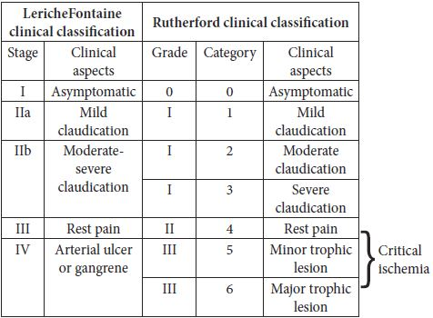
Acute and Chronic Limb Ischemia
Acute Ischemia
 (click to enlarge)
Chronic Ischemia
Acute Ischemia
- Pain
- Pallor
- Pulseless
- Parasthesia
- Paralysis
- Perishingly Cold
- Little to No Flow
Noninvasive Physiologic Testing for PAD
ABI
- ABI is an excellent screening test.
- Normal is >0.9 and <1.3
- Highest Ankle Pressure/Highest Brachial Pressure
- Excellent Screening Test:
- Surrogate for CAD
- Limitations
- Falsely elevated due to incompressibility from medial calcinosis
- DM, CKD (can use toe pressures in these patients instead).
ABI SCALE
>1.3 Calcified
0.9-1.3 Normal
0.5-0.9 Claudicant
<0.5 Critical Limb Ischemia
Segmental Limb Pressures
- Segmental pressures at upper/lower thigh, calf, ankle using PT/DP Doppler.
- Systolic pressure at upper thigh are 30-40 mmHg>brachial.
- Typically, a gradient of >20 mmHg between adjacent sides or between two sides at the same level is considered significant.
PVR (Air-filled Plethysmograph):
- Allows determination of location in arterial disease.
- Measures changes in volume of limb during cardiac cycles
- same cuffs as segmental pressures.
- Dependent on:
- cardiac pump, inflow vessels, tissue type/characteristics
- 1st finding is usually LOSS OF DICROTIC NOTCH.
- Advantages:
- Not affected by calcifications.
- Disadvantages:
- Can get “normalization” with collaterals
- Limited anatomic information.
Directional Doppler:
- Normal Waveform – Triphasic
- Abnormal – Monophasic
Segmental Doppler
Exercise Testing
TBI
- Normal:
- a toe pressure of 70 to 110 mmHg
- TBI > 0.5 to 0.75
- Diagnosis of Critical Limb Ischemia (CLI)
- Absolute Systolic Toe Pressure <30mmHg at Toe
- TBI < 0.2
- <50 mmHg: ischemic ulceration or gangrene
Other Tests:
- CLI:
- TBI TcPO2[transcutaneous oximetry test]
- SPP[skin perfusion pressure]
TcPO2 [transcutaneous oximetry] – measures oxygen tension 1-2 mm deep to the skin
- useful for wound healing prediction in extremities.
- Can be used to assess response to hyperbaric therapy.
- Low values seen in patients with PAD, CHF, edema, inflammation, callous or skin disease (scleroderma).
>70 mm Hg – Normal
<40 mm Hg – Impaired Wound Healing
<30 mm Hg – Critical Ischemia
SPP [skin perfusion pressure]
>50 mm Hg – Normal
40-50 mm Hg – Mild
30-40 mm Hg – Gray Area. Wound Healing Probable.
<30 mm Hg – Critical ischemia
CTA/MRA for PAD
- Mapping sites of stenosis or occlusion
- Analyzing best management plan (surgical or endovascular)
MR Angio :
- Advantages:
- No radiation.
- Disadvantages:
- Worse in setting of calcification
- Worse for assessment of arterial wall
CT Angio :
- Advantages:
- Better for assessment of calcification, artery wall.
- Disadvantages:
- Radiation, Iodine.
- Peripheral CTA requires good workstation and software.
- CPR[curved planar reformat]
- Advantages:
- Longitudinal cuts, Soft/hard plaque assessment
- Disadvantages:
- time consuming
- limited spatial resolution.
- Note: Infrapopliteal artery evaluation is limited in CTA
- use reconstruction filters, CPR, dual energy acquisition, bolus tracking.
- Advantages:
- CPR[curved planar reformat]
History & Physical for PAD (Highlights)
Ulcers Workup
History:
-What did the ulcer look like at first?
-When did it begin?
- Onset – Acute vs. Chronic
-Risk Factors:
- Has the patient had previous ulcers?
- Location previous ulcers, prior therapies
- Interventions/revascularizations, amputations?
-Family history of leg ulcers?
-Is the ulcer painful?
-What drugs/medications has the patient taken?
-History of other systemic disorders?
Past Medical History
-Does the patient have:
- PAD, venous disease or lymphedema
- DM, HTN, hyperlipidemia (TIA/Stroke or CAD)
- CKD (Calciphylaxis)
- Hematologic Disease (SCD, Thalassemia)
- Gastrointestinal Disease (pyoderma gangrenosum)
- Rheumatologic Disease (connective tissue disease)
- Skin Disease (psoriasis, tumors)
- Psychiatric or Neurologic Disease (factorial)
- New Medications (hydroxyurea)
Social or Family History
- History of tobacco/drug abuse?
- Occupation or prolonged history of standing?
- Does the patient have access to wound care?
- Can the patient do dressing changes (elderly or obese may have trouble reaching their lower legs)
- Psychological issues (factitial)?
- Family history of ulcers (metabolic/hematologic)?
Physical Exam
- Vital Signs (fever)
- Vascular Exam (presence of pulses)
- Color changes (pallor with elevation, dependent rubber)
- Edema
- Neuropathy
- Size (length, width, depth)
- Location, # of wounds
- Border – Induration
- Wound probe to bone
- Drainage (type, amount, color, odor)
- Painful
- Surrounding Skin (atrophic, thin, cellulitis)
LABS Routine
- CBC and differential
- UA, chemistry profile
- WSR, C-RP
- The WSR blood test is designed to either detect or monitor the presence of inflammation within the body.
- Thyroid Tests
- SPEP
- Albumin, Transferrin
Special Tests
- ANA, ANCA
- Complement
- Antiphospholipid antibodies and Thrombophilia screening
- Cyroglobulins, cold agglutinins, cryofibrinogen
Arterial Ulcers
- Painful at rest
- Nocturnal pain relieved by dependency
- Boney prominences, tips of toes, lateral malleolus, pre-tibial regions
- Ulcers usually sharply demarcated, punched out
- Base pale, gray, yellow dry +/- necrotic eschar
- Risk for major amputation if revascularization not performed
Venous Ulcers
- Occur over bony prominences – medial malleolus (gaiter area)
- Ulcer irregular, shallow with a beefy, red (granulation tissue), moist base
- Edema, hyperpigmentation, lipodermatosclerosis
- Occur spontaneously or from minor trauma
Neurotrophic Etiology – Ulcers
- Find on pressure points (plantar surfaces, medial/lateral metatarsal heads, base of the greater toe, posterior rim of the heel pad)
- Ulcer – deep and tracking surrounded by acute and chronic inflammation and callus formation
- Results from neuropathy, PAD and infection
- Painless
- Failure to heal may be due to inadequate offloading
Arterial and Venous (Mixed) Ulcers
- Under appreciated
- Unrecognized
- 15-20% of venous ulcers have PAD component
- Cardiovascular risk factors (present)
- Decreased pulses, painful, base fibrotic (check ACI)
- Treat arterial disease and add compression unless ABI <0.5 or the ankle pressure < 60 mmHg.
Treatment
- Recognize/treat underlying disease (accurate diagnosis)
- Arterial disease – perfusion evaluation – revascularization
- Venous disease – compression – control edema.
- Options:
- high strength elastic compression stockings, short stretch bandage, long stretch bandage, Unna’s paste boot, multilayer compressive wrap (elastic, inelastic), intermittent pneumatic compression.
- Options:
- Neuropathic disease – off-loading
- Infectious disease – infection control – antibiotics
- Wound debridement
- Maintain moist, clean environment for healing
- Minimize pain
- Recalcitrant ulcers – prolonged duration, large size
- consider adjunctive methods: endovascular, sclerotherapy (foam), surgery
- Prevention
Current PAD Prescription
- ASA 81 mg po qD OR Clopidogrel 75 mg po qD
- Tobacco Cessation Strategy
- Smoking Contributes to progression of disease, increased mortality, failure of revascularization.
- This should be discussed with each patient.
- Statin
- Lower LDL-C <100 mg/dL [or <70 mg/dL in high-risk patients]
- Simvastatin 40mg-80mg.
- Alternatively use Ezetimibe.
- Hyperlipidemia – All patients with PAD should be treated with a statin. Statin therapy improves CV and limb outcome
- IDEAL trial – high dose statin patients a/w with lower rates of PAD and lower coronary events
- Lower LDL-C <100 mg/dL [or <70 mg/dL in high-risk patients]
- Blood Pressure Reduction, prefer ACE-I <130/85
- Diabetes Therapy if HgbA1C>6
- Diabetes leads to increased progression of PAD, i.e. CLI.
- PAD patients with A1C<6.5 had lower rates of amputation compared to A1C>9.
- Important to have tight glycemic control.
- Exercise Prescription
- Foot Care Instructions
- Antiplatelet therapy
- (CAPRIE – RCT of ASA vs. Clopidogrel), CHARISMA).
- AHA guidelines recommend ASA or Clopidogrel in patients with PAD.
- CAPRIE trial:
- Clopidogrel is more effective than a medium dose (325 mg daily) aspirin in reducing serious vascular events and had a similar safety profile.
- (CAPRIE – RCT of ASA vs. Clopidogrel), CHARISMA).
- VOYAGER (RCT of Rivaraoxaban (Xarelto) + Aspirin vs. Aspirin alone)
- Demonstrate that Rivaroxaban 2.5 mg twice daily + ASA is associated with significantly lower incidence of composite outcome of acute limb ischemia, major amputation for vascular causes, myocardial infarction, ischemic stroke or death from cardiovascular causes than ASA alone.
- Cilastozol (Pletal)
- Improvement in symptoms with no change in CV associated death when compared to placebo
- Contraindicated in CHF. Although cilostazol appears to be safe for long-term administration, adherence is low (>60% discontinuation at 3 years) because of adverse effects
- including headache, palpitations, and diarrhea.
- Optimal dose of cilostazol is 100 mg twice daily, and it may take up to 4 months to derive maximum benefit from this drug.
- Pentoxyfiline: NOT recommended in treatment of PAD.
- Supervised Exercise Therapy – is beneficial in comparison to optimal medical therapy and should be considered prior to revascularization
- CLEVER study – better walking, improved symptoms with supervised walking